-
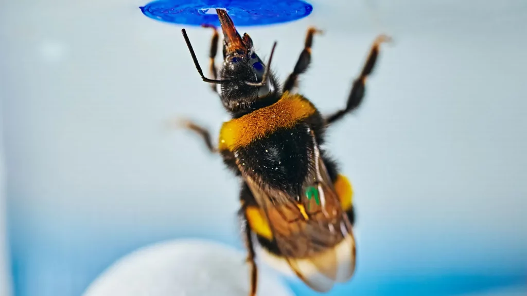 AI EditedThe headlines say bees can solve problems. The interesting part is how the researchers ruled out every boring explanation first... and how your students can practice the same thinking with a fish tank and a laser pointer.
 -
 AI Edited“How bad does it hurt?” It’s not for nothing that doctors usually struggle to ascertain our level of pain. It depends not only on how bad we report it to be, but also on the amount of pain we think we feel. But are there reasons behind it that would begin to decipher our (in)ability to […]
 -
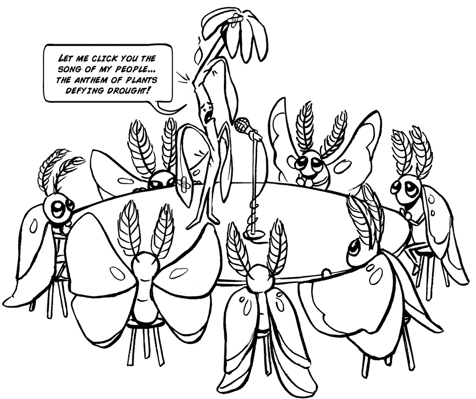 AI EditedIn a world of secrets, plants are speaking up. And science is all ears! As a recent study from the Cell journal shows, our leafy friends make popping or clicking sounds when under duress – such as when they are thirsty or injured. But how exactly do plants make sounds? A team of scientists from Tel […]
 -
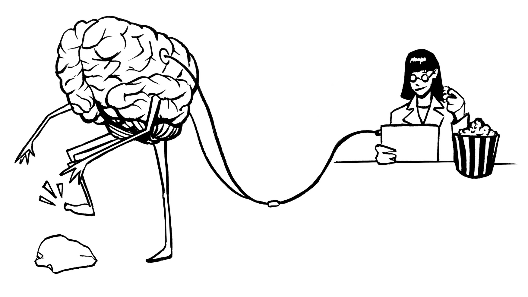
 AI EditedWhen it comes to the nervous system, you might think we’ve got the basics down. After all, it’s been over a century since the great Santiago Ramón y Cajal proposed the neuron doctrine, which basically said that the nervous system is made up of individual, discrete cells called neurons. As you may recall from […]